40 sponge anatomy diagram
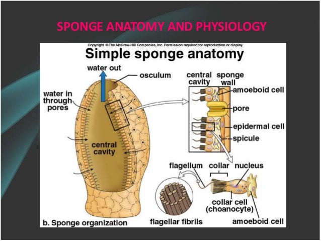
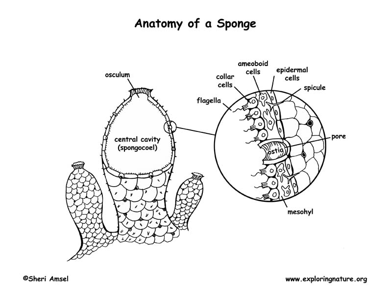
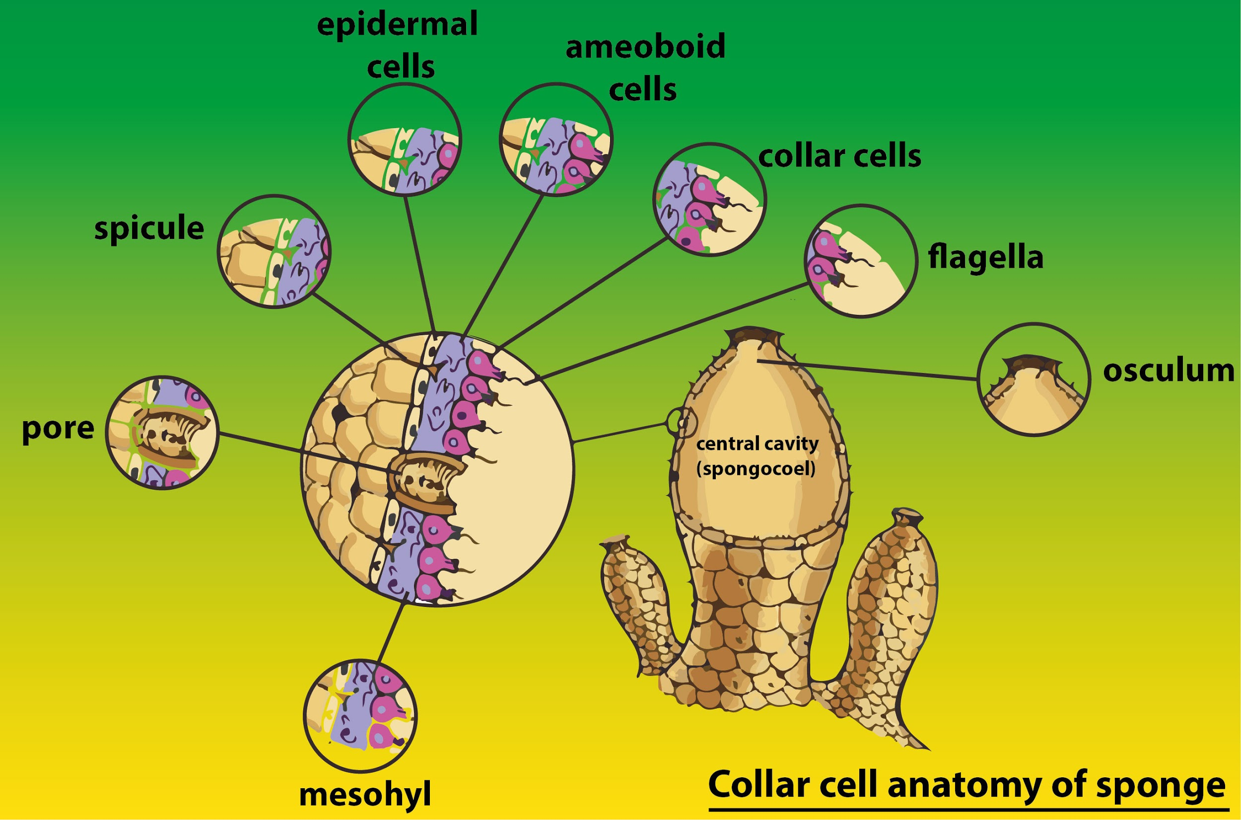
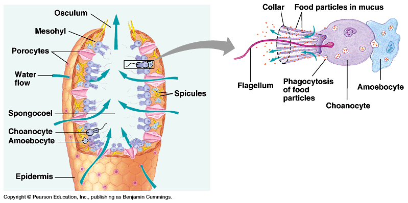
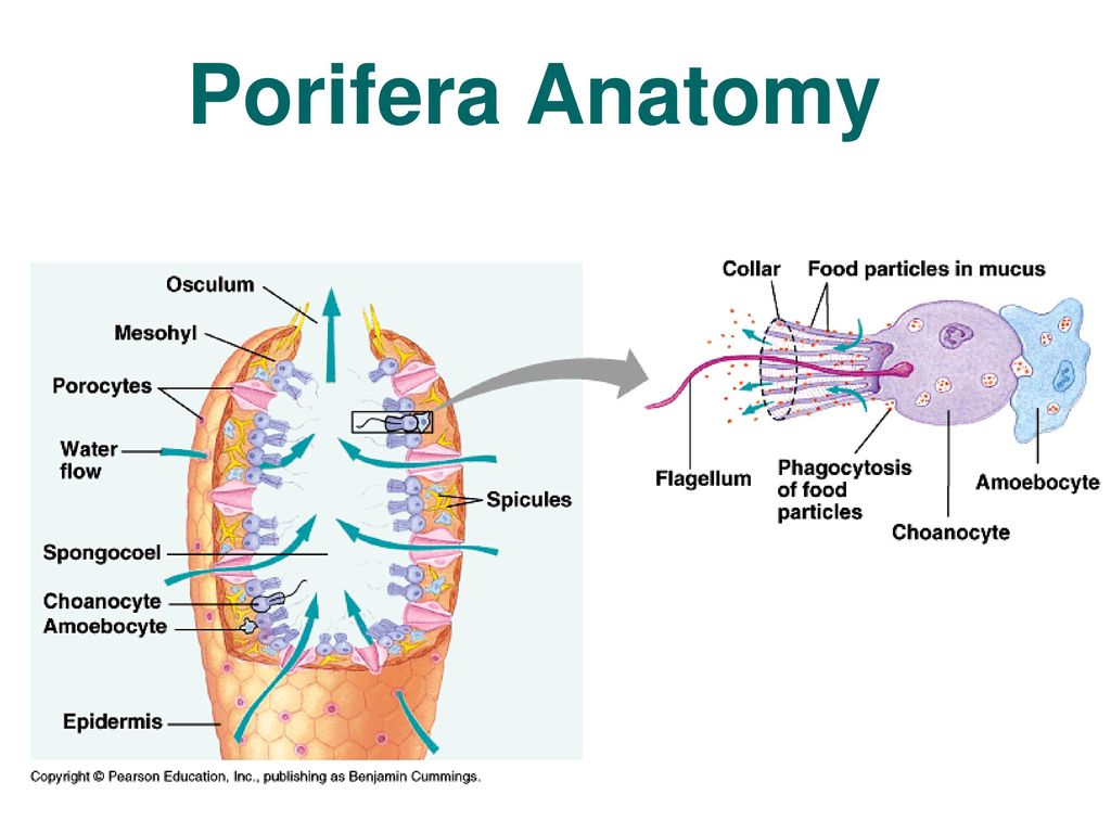
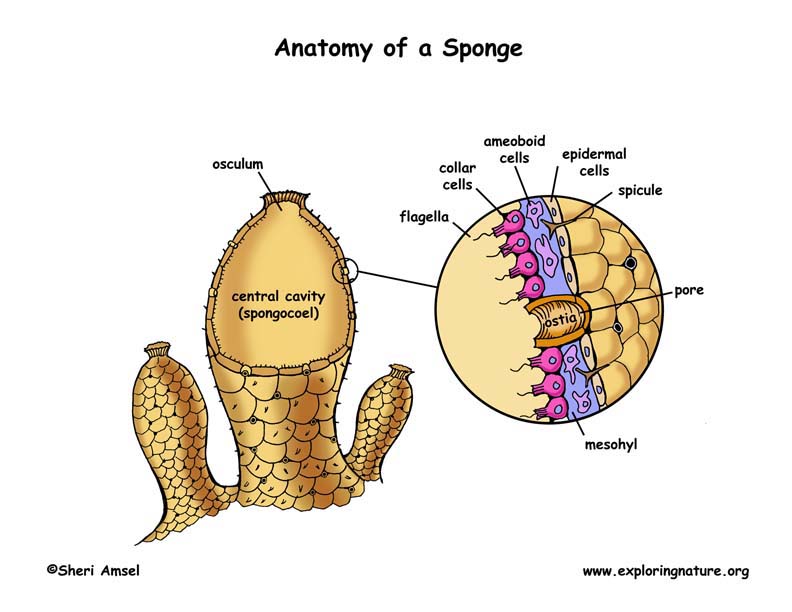
Sponge Anatomy Coloring Page - Exploring Nature Sponge Anatomy Coloring Page. PDF for Printing Out. Click Here. Link to More Info About this Animal (with Labeled Body Diagram) Click Here. Citing Research References. When you research information you must cite the reference. Citing for websites is different from citing from books, magazines and periodicals. The style of citing shown here is ... Solved Correctly label the following features of sponge ... Biology questions and answers. Correctly label the following features of sponge anatomy. I pore osculum w H.O out nucleus sponge wall spicule arnoebocyte epidermal cell H.O in through pores collar cell (choanocyte) amoebocyte collar central cavity flagellum < Prey 5 of 18 18 Next >. Question: Correctly label the following features of sponge ...
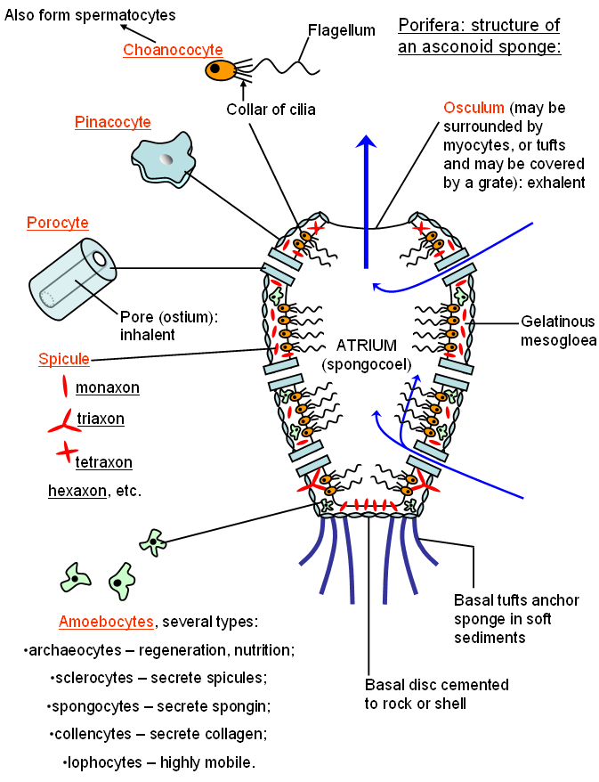
Modern sponge anatomy. ( A ) Schematic cross-section of ... Download scientific diagram | Modern sponge anatomy. ( A ) Schematic cross-section of simple asconoid sponge morphology with a central cavity and base. Pores (ostia) in body wall carry water ...
Sponge anatomy diagram
Clitoral complex — Anatomy Of Pleasure: Female The clitoral complex is an anatomical term (so don't worry if you haven't heard about it before) for the close relationship between the clitoris, the urethra or urethral sphincter and the low-down area at the base of the vagina.The urethral sphincter, sometimes known as the 'urethral sponge', is a tubular muscle that allows the urethra to open and close to let urine out and then close ... Sponges - EnchantedLearning.com Anatomy: The body of a sponge has two outer layers separated by an acellular (having no cells) gel layer called the mesohyl (also called the mesenchyme). In the gel layer are either spicules (supportive needles made of calcium carbonate) or spongin fibers (a flexible skeletal material made from protein). Sponges have neither tissues nor organs. Epithelium — Functions and Types of Epithelial Tissue | Lecturio Oct 05, 2020 · Definition of the Epithelial Tissue. The epithelium is a complex of specialized cellular organizations arranged into sheets without significant intercellular substance.They always occupy boundary surfaces of the body, i.e. they are located on the skin surface or the internal surface of hollow organs.
Sponge anatomy diagram. Shopping Online at Shopping.com | Price Comparison Site Shopping.com is a leading price comparison site that allows you to shop online for the best deals and lowest prices. Our mission is to help consumers use the power of information to easily find, compare and buy products online - in less time and for the best price! Structure of Sponges - Biodiscovery and the Great Barrier ... 03. SPONGES & CORALS 3(a) Structure of Sponges. Sponges are members of the Phylum Porifera (meaning 'pore-bearing') and are the oldest of all multi-cellular animals. They have been around for approximately 750 million years. They lack true tissues but have many cell types that take on these functions. Porifera: Anatomy - InfoPlease Porifera: Anatomy Sponges lack organs and tissue, and all the cells exhibit considerable independence. The sponge is made up of two single-cell-deep layers and an intermediate mesohyl (mobile cells plus extracellular matrix). The outer (sac) layer consists of flattened polygonal cells called pinacocytes. With Pleasure: A View of Whole Sexual Anatomy for Every ... Usually, when we're looking at a layout of sexual anatomy it's through the lens of reproduction, so it's all about penises and vaginas, testes and uterus.But from a standpoint of pleasure and sexual response, sexual anatomy is about far more than genitals and is far less about reproductive organs. Ultimately, all the parts and systems of the body are potential sexual organs in the context ...
Sponge - Wikipedia Sponges, the members of the phylum Porifera (/ p ə ˈ r ɪ f ər ə /; meaning 'pore bearer'), are a basal animal clade as a sister of the Diploblasts. They are multicellular organisms that have bodies full of pores and channels allowing water to circulate through them, consisting of jelly-like mesohyl sandwiched between two thin layers of cells.. Sponges have unspecialized cells that can ... Non-Subscriber Answer Page - EnchantedLearning.com Human Anatomy Mammals Plants Rainforests Sharks Whales. Physical Sciences: K-12 Astronomy The Earth Geology Hurricanes Landforms Oceans Tsunami Volcano: Languages ... The Anatomy Of Wood - Palomar College The microscopic cellular structure of wood, including annual rings and rays, produces the characteristic grain patterns in different species of trees.The grain pattern is also determined by the plane in which the logs are cut at the saw mill. Phylum - Porifera (Sponges) - Exploring Nature Body Traits (Anatomy): Sponges are made up of 2 body layers - an outer layer of epidermal cells and an inner layer of collar cells. The collar cells (choanocytes) trap food particles, while specialized ameboid cells (ameboctes) digest the particles and transport the nutrients to other cells throughout the sponge.
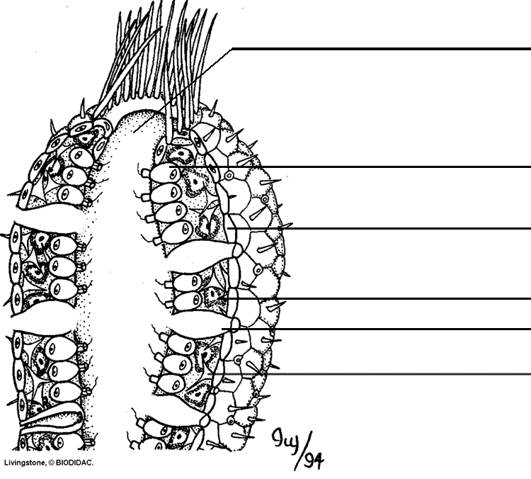
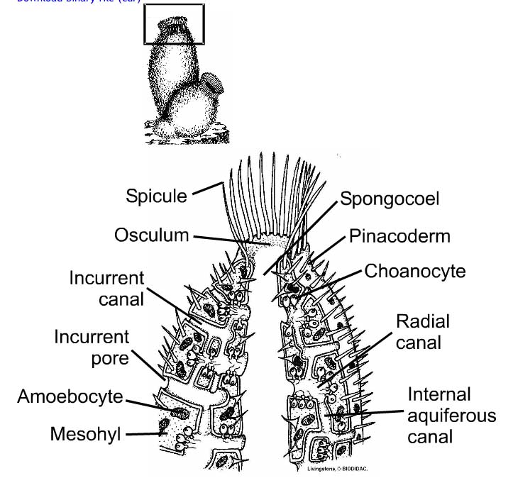
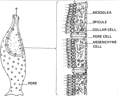
Sponge Anatomy Diagram Terms for labeling by Jacob ... Worksheet---Phylum Porifera - sponges; Pages 505-509 in Lab manual By Jacob Chiappetta Terms for Labeling the Sponge Anatomy Diagram , ( You may want to look at the slides in the folder- " Lab Practical # 2 Review - Phylum Animalia" or the Word bank for Practicum #2 Review to help label this diagram.) Identify the tissues, cells and regions on the sponge diagram that relate to the ... sponge - Form and function | Britannica sponge - sponge - Form and function: Sponges are unusual animals in that they lack definite organs to carry out their various functions. The most important structure is the system of canals and chambers, called a water-current system, through which water circulates to bring food and oxygen to the sponge. The water-current system also helps disperse gametes and larvae and remove wastes. Female reproductive system - Wikipedia The female reproductive system is made up of the internal and external sex organs that function in reproduction of new offspring.In humans, the female reproductive system is immature at birth and develops to maturity at puberty to be able to produce gametes, and to carry a foetus to full term. Scypha: History, Habitat and Nutrition (With Diagram) Scypha, also known as crown sponge, is a small, marine sponge found attached by a sticky secretion to some submerged solid object like rocks, shells of molluscs and corals. It is found in shallow water up to a depth of 50 fathoms (1 fathom = 6 feet) where waves provide the animal with plenty of food and well oxygenated water.
Sponge Anatomy - The Biology Corner Sponge Anatomy . Ostia | Osculum | Choanocyte | Ectoderm | Amebocyte | Spicule. This work is licensed under a Creative Commons Attribution-NonCommercial-ShareAlike 4.0 International License.Creative Commons Attribution-NonCommercial-ShareAlike 4.0 International License.
Sponge Anatomy Flashcards - Quizlet Start studying Sponge Anatomy. Learn vocabulary, terms, and more with flashcards, games, and other study tools.
Sponge Anatomy Quiz - PurposeGames.com About this Quiz. This is an online quiz called Sponge Anatomy. There is a printable worksheet available for download here so you can take the quiz with pen and paper.
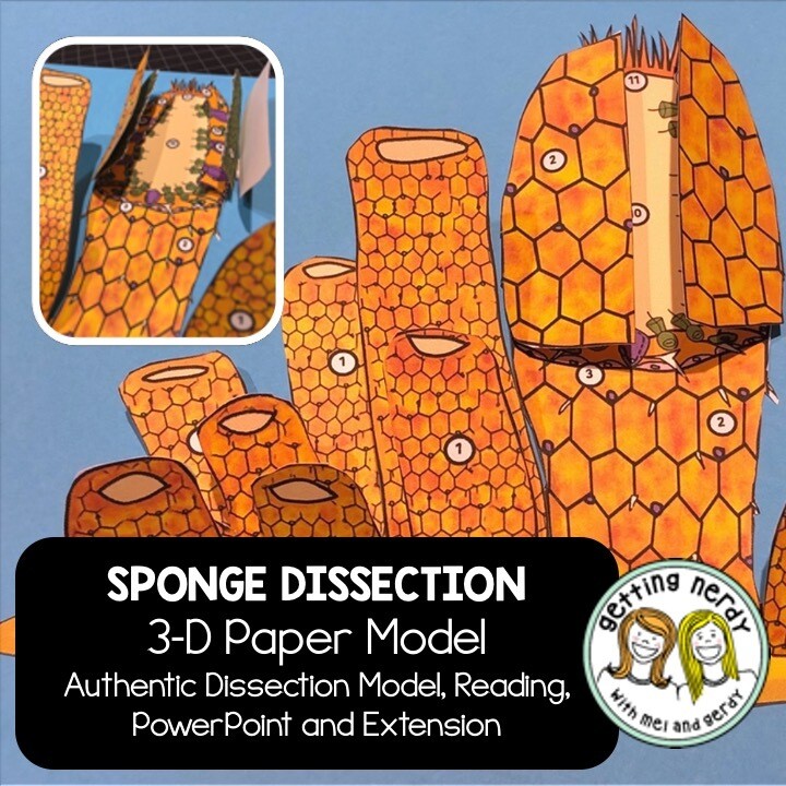
PDF 6 Sponge Dissection 2b Cnidaria (Jellyfish and Anemones) 9 ... 1. Remove a sponge from the jar, using forceps. Handle the sponge carefully, so as not to crush its internal support system. Sponges contain a skeleton that is composed of microscopic needles, called spicules. 2. Rinse the sponge gently under running water, to remove sea sand and any debris covering
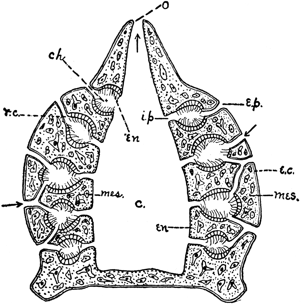
Sponges | ClipArt ETC "Diagram of simple type of sponge. c, cloaca; ch, chambers, lined with flagellate entoderm; e.p., external… Sponge The sponge, a many celled animal, begins its life as a single-cell, the egg.
Sponge Coloring - The Biology Corner Sponges belong to the kingdom animalia and the phylum porifera, though at one time they were thought to be plants. In this worksheet, read about sponges, and color a sponge to illustrate your knowledge of sponge anatomy.
Comparative Anatomy of Invertebrates Project - Mikayla ... Sponge *We didn't have a photo of the sponge dissection, but this is a diagram of one. Jellyfish. Oral Surface. Aboral Surface. ... Another thing that came through as fascinating to me was seeing the interior anatomy of the animals and how and where they functioned in the body. This assignment was definitely one of my favorites so far this year.
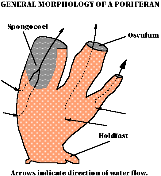
Label Sponge External Anatomy - EnchantedLearning.com Label Sponge External Anatomy Diagram Using the definitions listed below, label the sponge and the flow of water through it. A page on sponges epidermis - the layer of cells that covers the outer surface of the sponge. The thin, flattened cells of the epidermis are called pinacocytes. holdfast - root-like tendrils that attach the sponge to rocks.
Sponge Structure and Function - Advanced ( Read ... Sponge Structure and Function Sponges have three different body plans of sponges and use flagellated cells to pull seawater into their bodies to obtain particles of food. Progress
Porifera diagram | Biology notes, Science biology, Plant ... A beautiful Haliclona sp. specimen from Chek Jawa. Information about Porifera (Sponges) including their biology, anatomy, behaviour, reproduction, predators, prey and ecology. Jellyfish is free-swimming marine animal consisting of a gelatinous umbrella-shaped bell and trailing tentacles. Jellyfish are brainless, bloodless, b.
16 Best Images of Simple Microscope Labeling Worksheet ... Talking about Simple Microscope Labeling Worksheet, we've collected several similar images to complete your ideas. root diagram labeled, spinal cord cross section diagram labeled and sponge anatomy diagram answer key are three of main things we want to present to you based on the post title.
Study 13 Terms | Sponge Anatomy Diagram | Quizlet Start studying Sponge Anatomy. Learn vocabulary, terms, and more with flashcards, games, and other study tools.
Anatomy - The Basics OF: Porifera The Basics OF: Porifera Basic Anatomy Sponges contain no organs or even tissue. Instead, they consist of three cell sized layers. Compressed polygonal cells called pinacocytes make up the pinacoderm, the external sac layer. The cells in the outer layers can move inward and change function.
PDF Sponges A Coloring Worksheet Answer Key Sponges A Coloring Worksheet Answer Key Original Document: Sponges A Coloring Worksheet Students read about sponges, how they are classified, how they eat and where they are found. The instructions included specific information about sponge anatomy to be colored on diagram. Questions: 1.
Sponge Coloring Diagram and Questions - Biology Junction The primitive structure of a sponge consists of only two layers of cells separated by a non-living jelly like substance. The outer layer of the sponge is the epidermis which is made of flat cells called epithelial cells. Color all the epithelial cells (B) of the epidermis peach or pink.
Epithelium — Functions and Types of Epithelial Tissue | Lecturio Oct 05, 2020 · Definition of the Epithelial Tissue. The epithelium is a complex of specialized cellular organizations arranged into sheets without significant intercellular substance.They always occupy boundary surfaces of the body, i.e. they are located on the skin surface or the internal surface of hollow organs.
Sponges - EnchantedLearning.com Anatomy: The body of a sponge has two outer layers separated by an acellular (having no cells) gel layer called the mesohyl (also called the mesenchyme). In the gel layer are either spicules (supportive needles made of calcium carbonate) or spongin fibers (a flexible skeletal material made from protein). Sponges have neither tissues nor organs.
Clitoral complex — Anatomy Of Pleasure: Female The clitoral complex is an anatomical term (so don't worry if you haven't heard about it before) for the close relationship between the clitoris, the urethra or urethral sphincter and the low-down area at the base of the vagina.The urethral sphincter, sometimes known as the 'urethral sponge', is a tubular muscle that allows the urethra to open and close to let urine out and then close ...


















Comments
Post a Comment