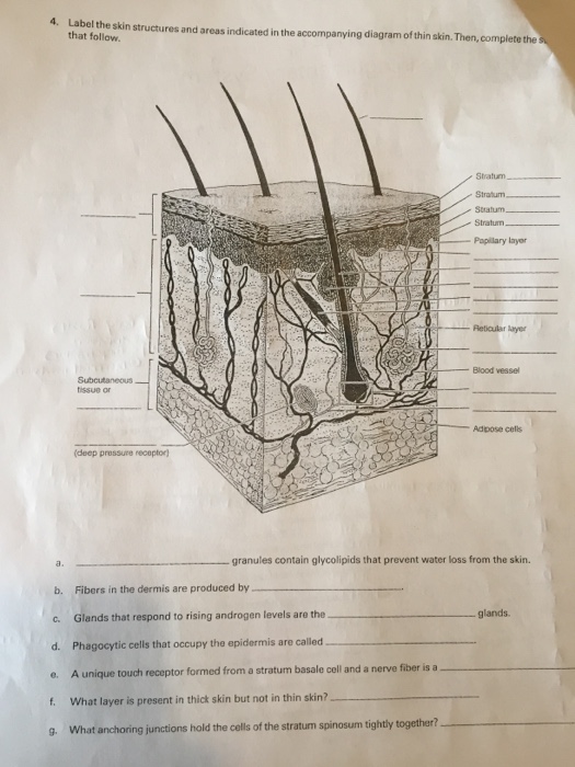
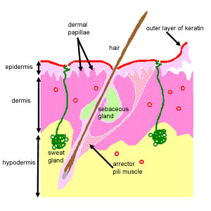
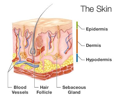
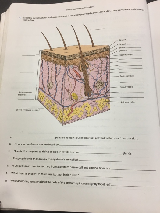
38 diagram of thin skin
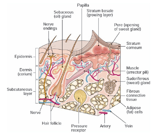
In the diagram of skin shown below, which structure illustrates that thin skin is present? a) C b) D c) E d) F e) H. a) a. In the diagram of skin shown above, which labeled structure generates fingerprints? a) A b) B c) G d) D. c) g. In the diagram of skin shown above, which area is primarily composed of areolar connective tissue? a) E b) F c) G Nov 16, 2018 · Diagram Of Thin Skin Structure. 5 1 Layers Of The Skin Anatomy And Physiology. Thick And Thin Skin Structure 14 Download Scientific Diagram. Solved Label The Skin Structures And Areas Indicated In The Ac. Skin Integumentary System.
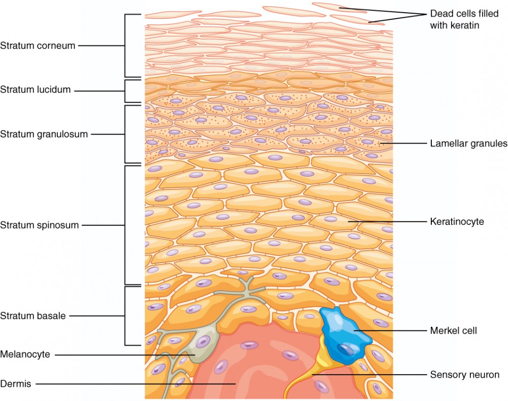
This thin layer of cells is found only in the thick skin of the palms, soles, and digits. The keratinocytes that compose the stratum lucidum are dead and flattened (see Figure 5.1.4 ). These cells are densely packed with eleiden , a clear protein rich in lipids, derived from keratohyalin, which gives these cells their transparent (i.e., lucid) appearance and provides a barrier to water.

Diagram of thin skin
AGING CHANGES. With aging, the outer skin layer (epidermis) thins, even though the number of cell layers remains unchanged. The number of pigment-containing cells (melanocytes) decreases. The remaining melanocytes increase in size. Aging skin looks thinner, paler, and clear (translucent). 3-D Skin Model Project - Anatomy and Physiology. This is a good basic project to have the students build a 3-D model of the skin. The project encourages students to build a 3-D Skin model to build, and describe the functions of various skin components. The assignment is out of 30 marks and a sample is attached on the second page. skin •Paper-thin skin •Dark or reddened areas 18 Darkly pigmented skin does not blanch. Parameter 3: Skin Color Redness •Reddened skin on the sacral area can be from a variety of etiologies. ... –Diagram of a body outline where staff can note any skin changes they observe. 30.
Diagram of thin skin. May 20, 2021 · Structure of thin skin of an animal. In the structure of the thin skin of animals, you will find the two distinct layers – the epidermis and dermis. I will show you the different histological characteristics from the epidermis and dermis of a skin microscope slide. #1. Sequential Easy First Hard First. Play as. Quiz Flashcard. In this quiz, we are going to focus on the skin structure of the human body. It’s easy to take your skin for granted, but when you consider how it protects your body from harm, it is something we should appreciate more. Do you know as much as you should about it? Questions and Answers. 1. Thin skin is a common condition in older adults, and is most noticeable in the face, arms, and hands. Treatment can prevent thin skin from getting worse. Anatomy of the Skin. The skin is a vital organ that covers the entire outside of the body, forming a protective barrier against pathogens and injuries from the environment. The skin is the body's largest organ; covering the entire outside of the body, it is about 2 mm thick and weighs approximately six pounds.
Layers of the Epidermis. Most areas of the body have four strata or layers. This is referred to as thin skin. In areas of the body exposed to greater friction, like the fingertips, palms and soles of the feet the epidermis has five strata or layers. This is referred to as thick skin. The epidermis is avascular (no blood vessels) and cells get their nutrients by way of diffusion from the deeper dermis (connective tissue). As cells move away from the dermis they start to dehydrate and die. In deserts, the human skin gets thicker to prevent water loss to dry air. Organisms with thin skin have the possibility of losing water all the time and need to stay near water to prevent it from drying. Sensation. Skin is the main sense organ that can sense touch, heat, pressure, cold, pain, and pleasure. Skin (Integumentary System) Blogger: Scientist. Thin Skin (Hematoxylin-Eosin) Thin Skin (Mallory Trichrome) Thin Skin (Verhoeff-Van Gieson) Differences between thick and thin skin in light microscope specimens. Share. skin •Paper-thin skin •Dark or reddened areas 18 Darkly pigmented skin does not blanch. Parameter 3: Skin Color Redness •Reddened skin on the sacral area can be from a variety of etiologies. ... –Diagram of a body outline where staff can note any skin changes they observe. 30.
3-D Skin Model Project - Anatomy and Physiology. This is a good basic project to have the students build a 3-D model of the skin. The project encourages students to build a 3-D Skin model to build, and describe the functions of various skin components. The assignment is out of 30 marks and a sample is attached on the second page. AGING CHANGES. With aging, the outer skin layer (epidermis) thins, even though the number of cell layers remains unchanged. The number of pigment-containing cells (melanocytes) decreases. The remaining melanocytes increase in size. Aging skin looks thinner, paler, and clear (translucent).
Copyright C The Mcgraw Hill Companies Chapter 18 Skin Skin Introduction The Skin Is The Largest Single Organ Of The Body Typically Accounting For 15 20 Of Total Body Weight And In Adults Presenting 1 5 2 M2 Of Surface To The External Environment

Sebaceous Gland Human Skin Sweat Gland Integumentary System Anatomy Biology Anatomy Human Anatomy Png Pngwing
:background_color(FFFFFF):format(jpeg)/images/library/13197/skin-histology_med_mag_new_mag_boxes_english.jpg)










Comments
Post a Comment